The electron microscopy (EM) laboratory team uses transmission electron microscopy (TEM), scanning electron microscopy (SEM), negative staining techniques and assays, immunogold labeling, quantitation of biological samples, and other specialized methods. The EM team at the Integrated Research Facility at Fort Detrick (IRF Frederick) uses these varied capabilities for the analysis of biological samples at an ultrastructural level.
Main Areas of Focus
The EM laboratory team focuses on the analysis and diagnosis of virus-infected tissues, cells, supernatants, and purified preparations.
Capabilities
- Custom protocols based on specific sample types and study goals
- Fresh chemical fixatives and custom reagents
- Precise protocols and methods for sample preparation
- Analysis and scientific evaluation of tissues, cells, and suspension samples
- Immunolabeling and virus quantitation assays
- High-resolution digital images from transmission and scanning electron microscopes
- Data interpretation and analysis
- Publication-quality data reports and analyses
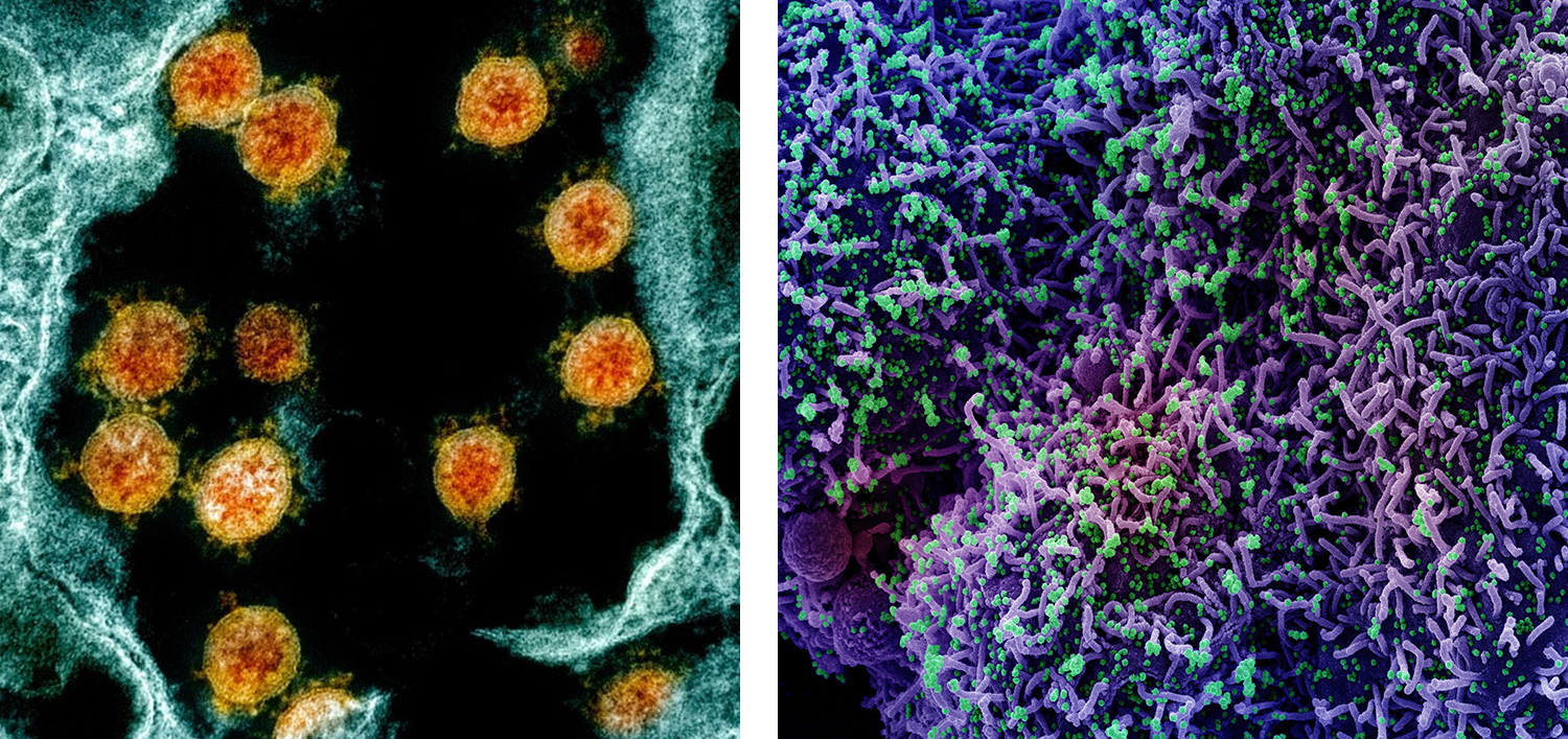
Left: Colorized transmission electron micrograph (TEM) of SARS-CoV-2 virus particles (orange) within an endosome of a Vero E6 cell (blue). Right: Colorized scanning electron micrograph (SEM) of CCL-81 cells (purple) infected with SARS-CoV-2 virus particles (green).
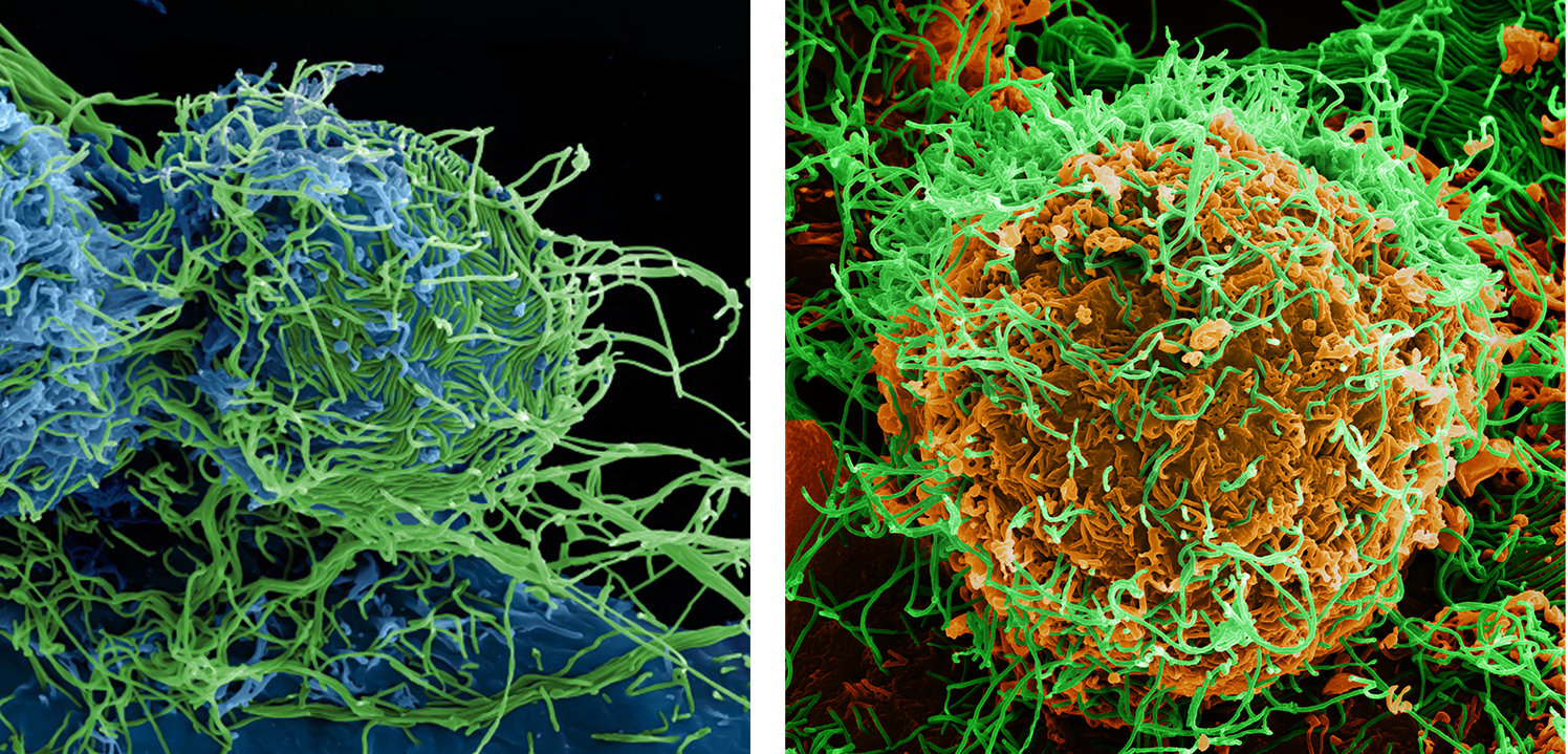
Left: Colorized scanning electron micrograph (SEM) of Vero E6 cells (blue) chronically infected with Ebola virus (EBOV) particles (green). Right: Colorized scanning electron micrograph (SEM) of a Vero E6 cell (orange) chronically infected with Ebola virus (EBOV) particles (green).
To explore an extensive gallery of images, including many generated through electron microscopy, visit NIAID’s Flickr account.
Location
Integrated Research Facility at Fort Detrick (IRF-Frederick)
Contact Information
Venkatesh Mani, Ph.D.
Director of Imaging and Maximum Containment (Contractor)
John Bernbaum
Electron Microscopist
IRF-Frederick
Standards
All procedures are well-documented and adhere to standard operating procedures (SOPs), methods, or study-approved plans and agreements.
Collaboration Opportunities
- Studies relevant to human disease
- Use of surrogate systems to test clinical hypotheses
- Use of biological systems to answer questions regarding disease pathogenesis and strategies for intervention including antimicrobials, vaccines, and other countermeasures
- Developing and incorporating cutting-edge technologies to understand infectious diseases
Read more about how to work with the IRF-Frederick.