Slideshow Block
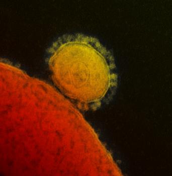
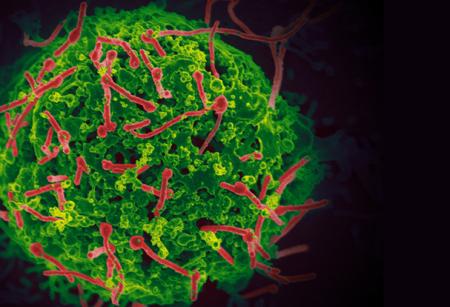
Transmission electron micrograph of Middle East respiratory syndrome coronavirus (MERS-CoV)
Credit
NIAID
he red staining visualizes the replication of MERS-CoV in the alveoli, the arrowheads indicate type I pneumocytes, and the arrows indicate type II pneumocytes
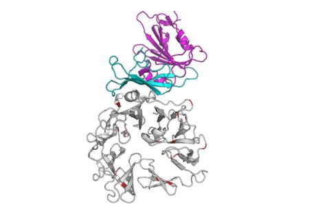
Graphic representation of the interaction between the MERS-CoV spike glycoprotein (blue-purple) and its receptor dipeptidyl peptidase 4 (DPP4).
Credit
NIAID
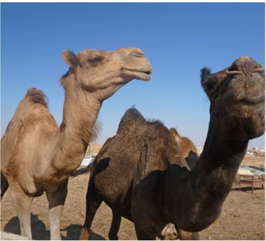
Dromedary camels were identified as the reservoir for MERS-CoV.
Credit
NIAID
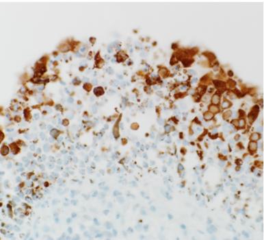
Replication of MERS-CoV in the cells lining the inside of the nose of dromedary camels
Credit
NIAID
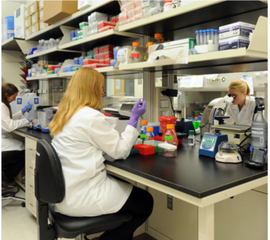
Biosafety level-2 (BSL-2) laboratory of the Virus Ecology Unit. From left to right: postbac Kerri Miazgowicz, Dr. Neeltje van Doremalen, and postbac Shauna Milne-Price.
Credit
NIAID
Ebola virus (red) budding from the surface of an infected cell.
Credit
NIAID
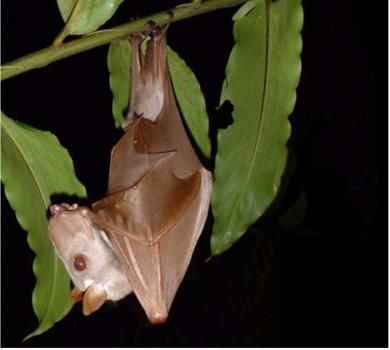
Male hammer-headed fruit bat (Hypsignathus monstrosus) in the Republic of the Congo. Hammer-headed fruit bats are followed over time to study the ecology of Ebola virus.
Credit
NIAID
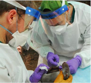
Obtaining a blood sample from a hammer-headed fruit bat for longitudinal Ebola virus ecology studies
Credit
NIAID
Biosafety level-2 (BSL-2) laboratory of the Virus Ecology Unit. From left to right: postbac Kerri Miazgowicz, Dr. Neeltje van Doremalen, and postbac Shauna Milne-Price.
Credit
NIAID
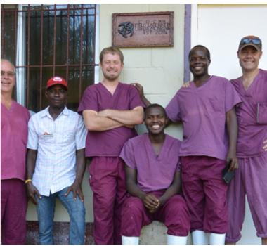
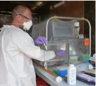
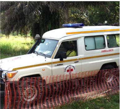
The Virus Ecology Unit working together with CDC-Kenya to provide Ebola virus diagnostics during the Ebola virus epidemic in West Africa