Ready, Set, Go—Immune System Status Predicts Future Responses

The Eyes Have it: A Functional Role for Prion Protein

NIAID Now |

In the early 1980s, scientists identified clumps of abnormal, misfolded prion protein in mammals as the cause of brain-wasting diseases, now called prion diseases. Since that time, they have struggled to answer: What does a normal prion protein do?

The answer, they believe, could help lead them to develop treatments and disease-prevention measures against human prion diseases, such as Creutzfeldt-Jakob disease, fatal familial insomnia and kuru, as well as animal prion diseases, such as scrapie in sheep and chronic wasting disease in cervids.

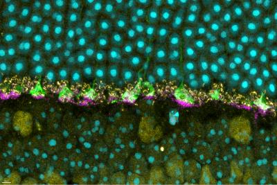
Now, a new study published in iScience from NIAID scientists at Rocky Mountain Laboratories in Hamilton, Montana, and colleagues provides details of how prion protein functions in the retina of mouse eyes, helping them respond to light.

The scientists used mice specially bred without prion protein to compare to wild mice with natural prion protein. Prior studies have suggested that prion protein may have a role in how nerves transmit signals to other nerves at specialized junctions, known as neural synapses. So, knowing that prion protein exists naturally in the eye, the researchers examined mouse retina for a specific neural synapse role.

A key tool the researchers used involved measuring the electroretinographic (ERG) responses – the amount of time it took for the retina in mice to respond to a flash of light. Remember as a kid in school learning about rods and cones in the eye and how they convert light signals to help the brain understand vision? The same is true in mice.

Compared to the wild mice with prion protein, the scientists observed deficiencies in ERG responses for mice without prion protein. The deficiencies affected the normal function of the rods and cones. And – using the ERG data and neural synapse information – they found that the deficiencies originated in the portion of the retina where natural prion protein was most highly concentrated.

Though additional study is needed, the researchers believe the prion protein may act like scaffolding to help cells and elements of the eye, such as rods and cones, to stabilize neural synapses. And they believe prion protein must be present for rods and cones to function normally.

The research team hopes these findings help colleagues who study prion diseases better understand what might occur in humans when natural forms of prion protein are therapeutically removed. New treatment strategies for prion diseases focus on using drugs that remove natural prion protein to eliminate the potential for misfolding and clumping. But researchers do not know whether that could result in unwanted outcomes, such as possibly affecting vision. These findings also could extend to other protein-related neurodegenerative diseases, such as Alzheimer’s (amyloid beta protein) and Parkinson’s diseases (alpha synuclein protein).

Scientists from Duke University and the McLaughlin Research Institute in Great Falls, Montana, collaborated on the study.

Reference: J Striebel, et al. The prion protein is required for normal responses to light stimuli by photoreceptors and bipolar cells. iScience DOI: 10.1016/j.isci.2024.110954 (2024).

Contact Information

Contact the NIAID Media Team.

301-402-1663
niaidnews@niaid.nih.gov

Search NIAID Blog

Household Bleach Inactivates Chronic Wasting Disease Prions

A 5-minute soak in a 40% solution of household bleach decontaminated stainless steel wires coated with chronic wasting disease (CWD) prions, . The scientists used the wires to model knives and saws that hunters and meat processors use when handling deer, elk and moose – all of which are susceptible to CWD.

Andre Ballesteros-Tato, Ph.D.

Section or Unit Name
Adaptive Immunity and Immunoregulation Section
Exclude from directory
Off
Section/Unit: Year Established
Section/Unit: Location
This Researcher/Clinician’s Person Page
Parent Lab/Program
Program Description

The overall goal of the Adaptive Immunity and Immunoregulation Section is to define the cellular and molecular mechanisms that regulate the balance between protective and pathogenic adaptive immune responses to allergens. Ultimately, our research aims to develop new immunotherapies to treat and prevent food and respiratory allergies without inducing profound immunosuppression.

We focus on three main areas:

  1. Tolerance vs. Inflammation: Tolerance prevents immune overactivation and maintains tissue homeostasis, while inflammation is critical for fighting infections. However, when these processes occur simultaneously, inflammation can disrupt tolerance, amplifying immune responses to harmless antigens such as allergens. Conversely, persistent allergic reactions can induce cellular and environmental changes that impair responses to pathogens and vaccines. We study how viral infections contribute to allergic responses and how allergies affect immune responses to pathogens. This knowledge is vital for designing therapies that prevent unwanted immune responses while preserving protective immunity.
  2. T Follicular Helper (Tfh) Cells: Tfh cells are crucial for supporting B cells and maintaining germinal centers. Recent findings from our lab have revealed that Tfh cells are more diverse than previously expected, secreting effector cytokines and playing broader regulatory roles. There is also growing evidence of an ontogenetic link between Tfh cells and other effector and regulatory T cell subsets. We study Tfh cell plasticity and heterogeneity, exploring their impact on tolerance induction and allergy development.
  3. Lung-Resident Memory T and B Cells: Lung-resident memory T and B cells are non-circulating memory cells that develop in response to respiratory challenges and permanently reside in the lungs. While the role of tissue-resident memory cells in response to respiratory pathogens has been established, their involvement in respiratory allergies remains elusive. We investigate how allergen-specific lung-resident memory T and B cells are generated and maintained, defining the factors controlling tissue memory generation and assessing their role in allergic responses. We also evaluate the potential of targeting these cells to prevent allergic reactions.

By integrating these projects, we aim to elucidate the complex mechanisms that balance protective and pathogenic immune responses and generate the necessary knowledge to develop novel treatments for food and respiratory allergies.

Selected Publications

Arroyo-Díaz NM, Bachus H, Papillion A, Randall TD, Akther J, Rosenberg AF, León B, Ballesteros-Tato A. Interferon-γ production by Tfh cells is required for CXCR3+ pre-memory B cell differentiation and subsequent lung-resident memory B cell responses. Immunity. 2023 Oct 10;56(10):2358-2372.e5.

Jenkins MM, Bachus H, Botta D, Schultz MD, Rosenberg AF, León B, Ballesteros-Tato A. Lung dendritic cells migrate to the spleen to prime long-lived TCF1hi memory CD8+ T cell precursors after influenza infection. Sci Immunol. 2021 Sep 10;6(63):eabg6895.

León B, Ballesteros-Tato A. Modulating Th2 Cell Immunity for the Treatment of Asthma. Front Immunol. 2021 Feb 10;12:637948.

Papillion A, Powell MD, Chisolm DA, Bachus H, Fuller MJ, Weinmann AS, Villarino A, O'Shea JJ, León B, Oestreich KJ, Ballesteros-Tato A. Inhibition of IL-2 responsiveness by IL-6 is required for the generation of GC-TFH cells. Sci Immunol. 2019 Sep 13;4(39):eaaw7636.

Botta D, Fuller MJ, Marquez-Lago TT, Bachus H, Bradley JE, Weinmann AS, Zajac AJ, Randall TD, Lund FE, León B, Ballesteros-Tato A. Dynamic regulation of T follicular regulatory cell responses by interleukin 2 during influenza infection. Nat Immunol. 2017 Nov;18(11):1249-1260.

León B, Bradley JE, Lund FE, Randall TD, Ballesteros-Tato A. FoxP3+ regulatory T cells promote influenza-specific Tfh responses by controlling IL-2 availability. Nat Commun. 2014 Mar 17;5:3495.

Visit PubMed for a complete publications listing.

Major Areas of Research
  • Characterize the mechanisms controlling adaptive immune responses, particularly memory T and B cells and T follicular helper cells, in the context of food and respiratory allergies
  • Investigate how infections contribute to the development of allergies and how allergies, in turn, affect immune responses to pathogens and vaccines
  • Develop novel immunotherapies that balance protection and immunosuppression for food and respiratory allergens

Andre Ballesteros-Tato, Ph.D.

Education:

Ph.D., Molecular Biology, 2007, Autónoma University of Madrid
B.S., Biology, 2001, University of Vigo

Languages Spoken: Spanish, Galician
Headshot of Andre Ballesteros-Tato.

Beatriz León, Ph.D.

Section or Unit Name
Innate Cells and Th2 Immunity Section
Exclude from directory
Off
Section/Unit: Year Established
Section/Unit: Location
This Researcher/Clinician’s Person Page
Parent Lab/Program
Program Description

Allergic diseases such as allergic rhinitis, asthma, and atopic dermatitis are characterized by an exaggerated immune response to otherwise harmless environmental proteins found in pollen, house dust mites, mold, cockroach debris, and pet dander. The immune system’s failure to maintain tolerance towards these allergens triggers a cascade of immune events, leading to chronic inflammation and tissue damage.

At the heart of allergic pathology is the intricate interaction between innate and adaptive immune cells, which coordinates the body's response to allergens. Key players in this process are T-helper type 2 (Th2) cells, a subset of T cells that orchestrate many of the immune mechanisms driving allergic inflammation.

Upon exposure to allergens, dendritic cells capture and process allergen-derived antigens, presenting them to naïve T cells in lymphoid tissues. In genetically or environmentally susceptible individuals, these naïve T cells differentiate into Th2 cells, which produce cytokines such as IL-4, IL-5, IL-9, and IL-13. These T cell-derived cytokines promote the production of IgE antibodies by B cells, sensitizing mast cells and basophils to allergens.

Additionally, these cytokines induce the activation and recruitment of eosinophils. Mast cells, basophils, and eosinophils then release mediators like histamine and proteases, leading to inflammation and allergic symptoms. Moreover, Th2 cells maintain a feedback loop that perpetuates chronic inflammation, contributing to conditions such as asthma, allergic rhinitis, and atopic dermatitis. Understanding the underlying immune mechanisms that lead to Th2 responses and their maintenance is crucial for developing novel therapeutic strategies to prevent and treat allergic conditions.

Our research team is dedicated to uncovering the fundamental mechanisms of airway and cutaneous allergic inflammation, primarily using mouse models. We focus on understanding how environmental allergens trigger and sustain allergic diseases, with particular attention to interactions between innate immune cells—such as monocytes, macrophages, and dendritic cells—and adaptive immune responses, especially Th2 cells. We explore how these immune interactions are influenced by the nature of allergens, environmental exposures, genetic factors, and microbiota.

Additionally, we investigate how these processes vary during sensitive periods, such as infancy and pregnancy, to better understand the onset and persistence of allergic inflammation. To advance our knowledge, we utilize advanced techniques, including conditional knockout murine models, multi-color flow cytometry, histology, functional lung assessment, microscopy, RNA-Seq, and single-cell technologies. Our ultimate goal is to identify targets for preventing or treating human allergic diseases.

Selected Publications

León B. A model of Th2 differentiation based on polarizing cytokine repression. Trends Immunol. 2023 Jun;44(6):399-407.

Bachus H, McLaughlin E, Lewis C, Papillion AM, Benveniste EN, Hill DD, Rosenberg AF, Ballesteros-Tato A, León B. IL-6 prevents Th2 cell polarization by promoting SOCS3-dependent suppression of IL-2 signaling. Cell Mol Immunol. 2023 Jun;20(6):651-665.

Kaur K, Bachus H, Lewis C, Papillion AM, Rosenberg AF, Ballesteros-Tato A, León B. GM-CSF production by non-classical monocytes controls antagonistic LPS-driven functions in allergic inflammation. Cell Rep. 2021 Dec 28;37(13):110178.

León B, Ballesteros-Tato A. Modulating Th2 Cell Immunity for the Treatment of Asthma. Front Immunol. 2021 Feb 10;12:637948.

Bachus H, Kaur K, Papillion AM, Marquez-Lago TT, Yu Z, Ballesteros-Tato A, Matalon S, León B. Impaired Tumor-Necrosis-Factor-α-driven Dendritic Cell Activation Limits Lipopolysaccharide-Induced Protection from Allergic Inflammation in Infants. Immunity. 2019 Jan 15;50(1):225-240.e4.

Ballesteros-Tato A, Randall TD, Lund FE, Spolski R, Leonard WJ, León B. T Follicular Helper Cell Plasticity Shapes Pathogenic T Helper 2 Cell-Mediated Immunity to Inhaled House Dust Mite. Immunity. 2016 Feb 16;44(2):259-73.

Visit PubMed for a complete publication listing.

Major Areas of Research
  • Immune sensing of environmental allergens
  • Innate and adaptive immune Interactions in T-helper type 2 (Th2)-driven allergy 
  • Environmental and genetic influences on type 2 inflammation

Beatriz León, Ph.D.

Education:

Ph.D., Molecular Biosciences, 2007, Autónoma University, Madrid, Spain
B.S., Biology, 2002, Complutense University, Madrid, Spain

Languages Spoken: Spanish
Headshot of Beatriz León.

Apply for Intramural NIAID Research Opportunities 2025

Candidate Malaria Vaccine Provides Lasting Protection in NIH-Sponsored Trials

Two National Institutes of Health (NIH)-supported trials of an experimental malaria vaccine in healthy Malian adults found that all three tested regimens were safe. One of the trials enrolled 300 healthy women ages 18 to 38 years who anticipated becoming pregnant soon after immunization. That trial began with drug treatment to remove malaria parasites, followed by three injections spaced over a month of either saline placebo or the investigational vaccine at one of two dosages.

Contact

Submit a Media Request

Contact the NIAID News & Science Writing Branch.

301-402-1663
niaidnews@niaid.nih.gov
All Media Contacts

Adriana A. de Jesus, M.D., Ph.D.

Section or Unit Name
Translational Autoinflammatory Diseases Section
Exclude from directory
Off
Section/Unit: Location
This Researcher/Clinician’s Person Page
Program Description

Dr. Adriana de Jesus is an assistant research physician in pediatric rheumatology translational research, particularly studying the genetic basis and pathogenic mechanisms of systemic autoinflammatory diseases (SAIDs).

She began investigating the genetic causes of autoinflammatory diseases while working on her Ph.D. in Brazil. She collaborated with the Translational Autoinflammatory Diseases Section (TADS) at NIH to report the first two Brazilian patients with a deficiency of interleukin 1 receptor antagonist (DIRA).

She also led a multicenter study for the genetic investigation of 102 patients with clinically suspected autoinflammatory diseases in Brazil. Since joining TADS in 2012, under the leadership of Dr. Raphaela Goldbach-Mansky, she has been responsible for the genetic investigation of patients with undifferentiated SAIDs enrolled into an NIH natural history protocol who undergo trio exome or genome sequencing.

Dr. Adriana de Jesus’ role in TADS involves generating and interpreting genetic data and playing a key part in the phenotypic characterizations of the patient cohorts. At TADS, she identified de novo variants in STING1 as the cause of STING-associated vasculopathy with onset in infancy (SAVI), allowing these patients to be treated with interferon-blocking therapies. She also identified de novo variants in NLRC4 and CDC42 as the genetic defect in two novel autoinflammatory diseases associated with macrophage activation syndrome (MAS).

Other significant contributions included the identification of variants in PSMG2 and PSMA5 in patients with proteasome-associated autoinflammatory syndromes (PRAAS), and IKBKG splice-site variants and SAMD9L frame-shift variants in patients with NEMO-deleted exon 5 autoinflammatory syndrome (NEMO-NDAS) and SAMD9L-associated autoinflammatory disease (SAAD), respectively.

She has also described a novel SAID caused by gain-of-function variants in LYN and characterized by skin vasculitis and liver fibrosis. Her contributions to the research on SAIDs also extend to the development and validation of a Type I interferon score assay, which is used as a biomarker to aid in the diagnosis and assessment of treatment responses in patients with Type I interferonopathies.

Selected Publications

de Jesus AA, Chen G, Yang D, Brdicka T, Ruth NM, Bennin D, Cebecauerova D, Malcova H, Freeman H, Martin N, Svojgr K, Passo MH, Bhuyan F, Alehashemi S, Rastegar AT, Uss K, Kardava L, Marrero B, Duric I, Omoyinmi E, Peldova P, Lee CR, Kleiner DE, Hadigan CM, Hewitt SM, Pittaluga S, Carmona-Rivera C, Calvo KR, Shah N, Balascakova M, Fink DL, Kotalova R, Parackova Z, Peterkova L, Kuzilkova D, Campr V, Sramkova L, Biancotto A, Brooks SR, Manes C, Meffre E, Harper RL, Kuehn H, Kaplan MJ, Brogan P, Rosenzweig SD, Merchant M, Deng Z, Huttenlocher A, Moir SL, Kuhns DB, Boehm M, Skvarova Kramarzova K, Goldbach-Mansky R. Constitutively active Lyn kinase causes a cutaneous small vessel vasculitis and liver fibrosis syndrome. Nat Commun. 2023 Mar 17;14(1):1502.

de Jesus AA, Hou Y, Brooks S, Malle L, Biancotto A, Huang Y, Calvo KR, Marrero B, Moir S, Oler AJ, Deng Z, Montealegre Sanchez GA, Ahmed A, Allenspach E, Arabshahi B, Behrens E, Benseler S, Bezrodnik L, Bout-Tabaku S, Brescia AC, Brown D, Burnham JM, Caldirola MS, Carrasco R, Chan AY, Cimaz R, Dancey P, Dare J, DeGuzman M, Dimitriades V, Ferguson I, Ferguson P, Finn L, Gattorno M, Grom AA, Hanson EP, Hashkes PJ, Hedrich CM, Herzog R, Horneff G, Jerath R, Kessler E, Kim H, Kingsbury DJ, Laxer RM, Lee PY, Lee-Kirsch MA, Lewandowski L, Li S, Lilleby V, Mammadova V, Moorthy LN, Nasrullayeva G, O'Neil KM, Onel K, Ozen S, Pan N, Pillet P, Piotto DG, Punaro MG, Reiff A, Reinhardt A, Rider LG, Rivas-Chacon R, Ronis T, Rösen-Wolff A, Roth J, Ruth NM, Rygg M, Schmeling H, Schulert G, Scott C, Seminario G, Shulman A, Sivaraman V, Son MB, Stepanovskiy Y, Stringer E, Taber S, Terreri MT, Tifft C, Torgerson T, Tosi L, Van Royen-Kerkhof A, Wampler Muskardin T, Canna SW, Goldbach-Mansky R. Distinct interferon signatures and cytokine patterns define additional systemic autoinflammatory diseases. J Clin Invest. 2020 Apr 1;130(4):1669-1682.

de Jesus AA, Brehm A, VanTries R, Pillet P, Parentelli AS, Montealegre Sanchez GA, Deng Z, Paut IK, Goldbach-Mansky R, Krüger E. Novel proteasome assembly chaperone mutations in PSMG2/PAC2 cause the autoinflammatory interferonopathy CANDLE/PRAAS4. J Allergy Clin Immunol. 2019;143(5):1939-1943.

Canna SW, de Jesus AA, Gouni S, Brooks SR, Marrero B, Liu Y, DiMattia MA, Zaal KJ, Sanchez GA, Kim H, Chapelle D, Plass N, Huang Y, Villarino AV, Biancotto A, Fleisher TA, Duncan JA, O'Shea JJ, Benseler S, Grom A, Deng Z, Laxer RM, Goldbach-Mansky R. An activating NLRC4 inflammasome mutation causes autoinflammation with recurrent macrophage activation syndrome. Nat Genet. 2014 Oct;46(10):1140-6.

Liu Y, Jesus AA, Marrero B, Yang D, Ramsey SE, Sanchez GAM, Tenbrock K, Wittkowski H, Jones OY, Kuehn HS, Lee CR, DiMattia MA, Cowen EW, Gonzalez B, Palmer I, DiGiovanna JJ, Biancotto A, Kim H, Tsai WL, Trier AM, Huang Y, Stone DL, Hill S, Kim HJ, St Hilaire C, Gurprasad S, Plass N, Chapelle D, Horkayne-Szakaly I, Foell D, Barysenka A, Candotti F, Holland SM, Hughes JD, Mehmet H, Issekutz AC, Raffeld M, McElwee J, Fontana JR, Minniti CP, Moir S, Kastner DL, Gadina M, Steven AC, Wingfield PT, Brooks SR, Rosenzweig SD, Fleisher TA, Deng Z, Boehm M, Paller AS, Goldbach-Mansky R. Activated STING in a vascular and pulmonary syndrome. N Engl J Med. 2014 Aug 7;371(6):507-518.

Jesus AA, Osman M, Silva CA, Kim PW, Pham TH, Gadina M, Yang B, Bertola DR, Carneiro-Sampaio M, Ferguson PJ, Renshaw BR, Schooley K, Brown M, Al-Dosari A, Al-Alami J, Sims JE, Goldbach-Mansky R, El-Shanti H. A novel mutation of IL1RN in the deficiency of interleukin-1 receptor antagonist syndrome: description of two unrelated cases from Brazil. Arthritis Rheum. 2011 Dec;63(12):4007-17.

Visit Dr. Adriana de Jesus’ complete publication list.

Major Areas of Research
  • Identify genetic causes of undifferentiated systemic autoinflammatory diseases by next-generation sequencing
  • Identify genetic diagnoses in patients affected by early onset Type I interferon (IFN)-mediated autoinflammatory diseases (interferonopathies)
  • Study the pathogenesis of Type I interferonopathies, including proteasome-associated autoinflammatory syndromes (PRAAS), stimulator of interferon genes (STING)-associated vasculopathy with onset in infancy (SAVI), and undifferentiated Type I interferonopathies
  • Diagnose and treat patients with systemic autoinflammatory diseases