A Secret to Health and Long Life? Immune Resilience, NIAID Grantees Report

NIAID Now |

Do you know some people who almost never get sick and bounce back quickly when they do, while other people frequently suffer from one illness or another? NIAID-supported researchers have pinpointed an attribute of the immune system called immune resilience that helps explain why some people live longer and healthier lives than others.

Immune resilience involves the ability at any age to control inflammation and to preserve or rapidly restore immune activity that promotes resistance to disease, the investigators explain. They discovered that people with the highest level of immune resilience lived longer than others. People with greater immune resilience also were more likely to survive COVID-19 and sepsis as well as to have a lower risk of acquiring HIV infection and developing AIDS, symptomatic influenza, and recurrent skin cancer. In addition, women were more likely to have optimal immune resilience than men. These findings suggest that knowing an individual’s level of immune resilience could help healthcare providers assess the risk for a severe outcome in people with immunity-dependent diseases and identify mechanisms to extend lifespan, according to the investigators. The NIAID co-funded research was published today in the journal Nature Communications.

The nine-year study was led by Sunil Ahuja, M.D., the President's Council/Dielmann Chair for Excellence in Medical Research and professor of medicine at the University of Texas Health Science Center at San Antonio. Dr. Ahuja is also director of research enhancement programs at the university and director of the Veterans Administration Center for Personalized Medicine in the South Texas Veterans Health Care System in San Antonio.

Measuring Immune Resilience

Dr. Ahuja and colleagues developed two ways to measure immune resilience, or IR, one based on immune-cell levels in blood and the other on patterns of genes that are turned on, or expressed. The investigators evaluated these metrics in roughly 48,500 people ages 9 to 103 years who were exposed to pathogens and other immune-system stressors of varied types and severity levels, including the natural aging process. The data on these people, who were Black, Hispanic, or White, came from more than 18 different studies conducted in Africa, Europe and North America.

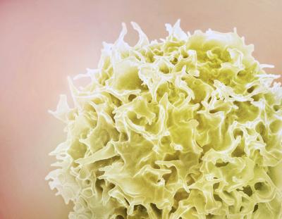
One of the two IR metrics—the immune health grade, or IHG—is based on the relative quantities of two types of white blood cells, CD8+ T cells and CD4+ T cells, which coordinate the immune system’s response to pathogens and kill other cells that have been infected. CD4+ T-cell counts in the blood have long been used to measure immune health, particularly in people with HIV. The IHG is innovative because it reflects the balance between CD8+ and CD4+ T-cell counts. The CD8+ to CD4+ T-cell balance in optimal IR is called IHG-I, while less optimal levels of IR are called IHG-II, IHG-III and IHG-IV.

The second IR metric is based on two patterns of gene expression: one that best predicted survival and another that best predicted death in two large groups of people after controlling for age and sex. The researchers labeled the survival-associated pattern SAS-1 and the mortality-associated pattern MAS-1. SAS-1 genes are largely related to immune competence—the ability to preserve or rapidly restore immune activity that promotes resistance to disease. MAS-1 genes are largely related to inflammation—the process by which the immune system recognizes and helps kill or remove pathogens and other harmful or foreign substances and begins the healing process. The scientists found that high levels of SAS-1 gene expression and low levels of MAS-1 gene expression indicated that a person had optimal IR and a lower risk of dying prematurely, while the opposite indicated poor IR and a higher risk of premature death. If SAS-1 and MAS-1 levels were both high or both low, IR and risk of premature death were moderate.

The investigators tested these two sets of metrics—IHGs and SAS-1/MAS-1—in the context of low-, moderate- and high-intensity stress to the immune system to determine how well the measures predicted health outcomes and lifespan after controlling for age and sex. The scientists identified groups of people experiencing these different intensities of immune challenges in the context of their daily lives. The group experiencing low-intensity immune stimulation comprised thousands of HIV-negative people ages 18 to 103 years participating in long-term studies of aging. The group experiencing moderate-intensity immune stimulation involved hundreds of HIV-negative people with SARS-CoV-2 infection, autoimmune disease, kidney transplant, or behavioral risk factors for acquiring HIV. Finally, the group experiencing high-intensity immune stimulation comprised thousands of people whose immune systems were responding to HIV replication in the blood soon after infection.

Variations in Immune Resilience

The researchers found that preserving optimal IR, as indicated by having either IHG-I or the combination of high SAS-1 and low MAS-1, was associated with the best health outcomes and longest lifespans. In addition, the risk or severity of negative immunity-dependent health outcomes increased as baseline IR level decreased. The scientists also demonstrated that the proportion of people with optimal IR (IHG-I or high SAS-1/low MAS-1) tended to be highest in the youngest people and lowest in the oldest people. Similarly, the proportion of people with the least optimal IR metrics (IHG-III or IHG-IV and low SAS-1/high MAS-1) tended to be lowest in the youngest age groups and highest in the oldest age groups. However, the investigators found that each of the four immune health grades and related SAS-1/MAS-1 gene expression profiles appeared in people in every age group.

As people age, the researchers explained, increasingly more health conditions such as acute infections, chronic diseases and cancers challenge their immune systems to respond and—ideally—recover. Over time, these challenges degrade most people’s immune health, accounting for the declining proportion of people with IHG-I and high SAS-1/low MAS-1 over the lifespan. However, some people who are 90 years old or more still have IHG-I and high SAS-1/low MAS-1—a reflection of their immune systems’ exceptional capacity to control inflammation and preserve or rapidly restore immune activity associated with longevity despite the many immune health challenges they have faced.

By contrast, the researchers demonstrate that some young adults who are repeatedly exposed to immune threats may have the least optimal IR, as measured by IHG-III or IHG-IV and low SAS-1/high MAS-1. The investigators show how young female sex workers who had many clients and did not use condoms—and thus were repeatedly exposed to sexually transmitted pathogens—had drastically degraded immune health even if they did not acquire HIV. In addition, sex workers with nonoptimal IR, especially those with IHG-IV, had a higher risk of acquiring HIV infection regardless of their level of risk behavior. However, most of the sex workers who began reducing their exposure to sexually transmitted pathogens by using condoms and decreasing their number of sex partners improved to IHG-I over the next 10 years.

The scientists also observed this plasticity of IR in other contexts. For example, the researchers found that most people couldn’t maintain optimal IR when they experienced inflammatory stress from a common symptomatic viral infection like a cold or the flu. In this situation, most people who the investigators studied developed low SAS-1/high MAS-1 within 48 hours of symptom onset, indicating poor IR and a high risk of dying prematurely. As people recovered from their infection, however, many gradually returned to the more favorable SAS-1/MAS-1 levels that they had before. Yet nearly 30 percent of those who had high SAS-1/low MAS-1 before getting sick did not fully regain that survival-associated profile by the end of the cold and flu season, even though they had recovered from their illness.

Interestingly, the investigators also found that the ability to maintain or develop optimal IR during a respiratory virus infection, as measured by high SAS-1/low MAS-1, correlated with an absence of symptoms.

Implications of Immune Resilience

The researchers suggest numerous implications of their findings for personalized medicine, biomedical research, and public health. First, some younger adults have low IR due to unsuspected immunosuppression, whereas some older adults have superior IR. These differences may account for why some younger people are predisposed to disease and shorter lifespans while some elderly people remain unusually healthy and live longer than their peers. Second, reducing exposure to immune stressors may maintain optimal IR or give people with low or moderate IR the opportunity to regain optimal IR, thereby decreasing risk of severe disease. Third, measuring people’s IHG and SAS-1/MAS-1 profile in the early stages of illness could allow for detection of poor IR and initiation of more aggressive therapy. Fourth, it may make sense to balance the intervention and placebo arms of clinical trials by both IR status and common factors such as age and sex when testing interventions dependent on controlling inflammation and preserving or rapidly restoring immune activity associated with longevity. Fifth, developing and implementing strategies to mitigate IR degradation may improve people’s response to vaccination as well as their overall health and lifespan. Finally, strategies for boosting IR and reducing recurrent immune stressors may help address racial, ethnic, and geographic disparities in diseases such as cancer and viral infections like COVID-19.

Reference: SK Ahuja, et al. Immune resilience despite inflammatory stress promotes longevity and favorable health outcomes including resistance to infection. Nature Communications DOI: 10.1038/s41467-023-38238-6 (2023).

Contact Information

Contact the NIAID Media Team.

301-402-1663
niaidnews@niaid.nih.gov

Search NIAID Blog

NIAID-Supported Study Provides Stronger Evidence of Link Between RSV and Childhood Asthma

NIAID Now |

A NIAID-supported study has found that respiratory syncytial virus (RSV) infection in the first year of life is associated with a significantly increased risk of asthma in children. The study was published in the journal The Lancet. These findings provide additional evidence for a casual association between the occurrence of RSV infection in children younger than 1 and an increased incidence of wheezing and asthma in later in life.

RSV is a seasonal respiratory pathogen that affects almost all children by age 2, and repeatedly throughout life. In most children, symptoms of the virus are mild and usually resolve within a week. However, RSV can lead to death or serious illness, especially in premature or very young infants, and those with chronic lung disease or congenital heart disease, although about half of all RSV hospitalizations are among healthy infants. In infants (children younger than 1), RSV is the leading cause of bronchiolitis, a lower respiratory tract infection in infants and young children presenting with coughing and wheezing. Prior evidence on the links between RSV infection in infancy and respiratory health derives from studies of children with severe RSV bronchiolitis, which impacts a minority of children. The population-level asthma risk following RSV infection of any severity has not been studied before.

The “Infant Susceptibility to Pulmonary Infections and Asthma Following RSV Exposure (INSPIRE)” is the first cohort specifically designed to test the hypothesis that not being infected with RSV in infancy decreases the risk of childhood asthma. The population-based cohort study included 1,946 healthy infants born between June and December of 2012 and 2013 who were 6 months old or younger at the beginning of the RSV season (November to March in the study area of Tennessee). Biweekly surveillance and serology tests were used to classify infants as RSV infected or not infected in the first year of life. Of the 1,741 who received classifications, 54% were infected with RSV in the first year of life.

Participants were followed prospectively for five years and then evaluated for 5-year current asthma, which was defined as 1) a parental report of diagnosed asthma or the use of asthma medications before age 5, and 2) any of the following in the 12 months prior to the 5-year visit: asthma symptoms, use of systemic (oral or intravenous) steroids for asthma, or doctor or emergency room visits for asthma symptoms.

The study found that infants who were not infected with RSV in the first year of life had a 26% lower risk of asthma at 5 years of age than those who were infected with RSV as infants. Because the study was observational, the results do not definitively establish causality but do support a need for long-term follow-up of common respiratory outcomes among children in clinical trials of RSV prevention products.

Infancy is a critical time for immune system and pulmonary development; understanding how RSV infection before age 1 is associated with an increased risk of childhood asthma could help to prevent long-term childhood respiratory morbidity.

The study was funded by NIAID, the National Heart, Lung and Blood Institute, the National Center for Advancing Translational Sciences and the Eunice Kennedy Shriver National Institute of Child Health and Human Development.

Reference: C Rosas-Salazar, et al. Respiratory syncytial virus infection during infancy and asthma during childhood in the USA (INSPIRE): a population-based, prospective birth cohort study.

The Lancet DOI: 10.1016/S0140-6736(23)00811-5 (2023)

Contact Information

Contact the NIAID Media Team.

301-402-1663
niaidnews@niaid.nih.gov

Search NIAID Blog

Forgoing One Food Treats Eosinophilic Esophagitis as Well as Excluding Six

Eliminating animal milk alone from the diet of adults with eosinophilic esophagitis, or EoE, is as effective at treating the disease as eliminating animal milk plus five other common foods, a clinical trial funded by the National Institutes of Health has found. For people with EoE whose disease remains active after they forgo animal milk, a more restrictive diet may help them achieve remission, according to the researchers. These findings were published today in the journal The Lancet Gastroenterology & Hepatology

Contact

Submit a Media Request

Contact the NIAID News & Science Writing Branch.

301-402-1663
niaidnews@niaid.nih.gov
All Media Contacts

Some Reported Allergic Reactions to mRNA COVID-19 Vaccines Were Likely Stress Responses

NIAID Now |

NIAID Investigators Demonstrate How Stress Responses Closely Mimic Allergic Reactions

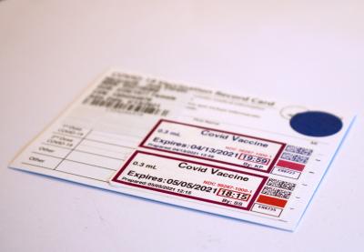
Some responses to the mRNA COVID-19 vaccines reported as severe allergic reactions were likely a recently described, non-allergic condition called immunization stress-related response (ISRR), according to NIAID investigators. The symptoms of ISRR can closely mimic those of a severe allergic reaction known as anaphylaxis, a potentially life-threatening, systemic allergic reaction in which the immune system releases a dangerous flood of chemicals. These findings from a small study are being presented today at the 2023 Annual Meeting of the American Academy of Allergy, Asthma & Immunology in San Antonio. 

As defined by the World Health Organization, ISRR encompasses a range of signs and symptoms that may arise before, during or immediately after immunization and are related to stress from the process of immunization, not to the vaccine product. Some of the manifestations of ISRR are the same as those that occur during anaphylaxis, including a rapid pulse, throat tightness, lightheadedness, trouble breathing, and nausea.

The reported rate of severe allergic reactions to immunization with mRNA COVID-19 vaccines, while estimated to be on the order of just five cases per million doses administered, is still higher than that reported for conventional vaccines. To better understand this phenomenon, NIAID researchers assessed the safety of a second dose of an mRNA COVID-19 vaccine in people who had experienced a systemic allergic reaction after their first dose. The study was led by Muhammad B. Khalid, M.D., a clinical fellow in the NIAID Laboratory of Allergic Diseases, and Pamela A. Frischmeyer-Guerrerio, M.D., Ph.D., the laboratory chief.

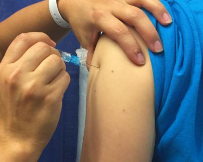
The investigators enrolled 16 people ages 16 to 69 years who had a moderate or severe systemic allergic reaction to their first dose of the Moderna or Pfizer-BioNTech COVID-19 vaccine. The study participants were admitted for a minimum of four days to the Intensive Care Unit at the NIH Clinical Center in a closely supervised, safe and controlled environment. There, they received the Pfizer-BioNTech mRNA COVID-19 vaccine and a look-alike injection of placebo in a random order on different days. Neither the participants nor the investigators knew which shot was being given on which day. At admission and during the inpatient stay, participants underwent breathing tests and frequent blood draws so medical staff could discern the details of any allergic or other responses to the vaccine.

People who had an allergic reaction to the first dose of a vaccine would be expected to have an allergic reaction to subsequent doses with equal or greater severity. However, only three participants, or 19%, developed an allergic reaction within minutes of receiving the second dose of vaccine, and one reaction was mild while two were moderate. As expected, no one developed an allergic reaction to the placebo.

By contrast, nine study participants developed a non-allergic reaction within minutes of receiving the vaccine, and 11 developed a non-allergic reaction within minutes of receiving the placebo. The non-allergic reactions included numbness, tingling, dizziness, throat tightness, difficulty swallowing, and temporarily high blood pressure—signs and symptoms consistent with ISRR. Forty-five percent of these reactions were classified as moderate to severe.

To further probe the nature of the volunteers’ responses to mRNA COVID-19 vaccines, the study team administered an open-label booster dose of the Pfizer-BioNTech vaccine and performed skin testing for allergic reactions. Thirteen participants completed this part of the study and another three were scheduled to do so.

The skin testing began with a so-called skin-prick test. A tiny drop of the Pfizer-BioNTech vaccine, a vaccine component called polyethylene glycol (PEG), and a related chemical called polysorbate were placed on the skin, and a small prick was made through each drop into the skin. If this did not elicit a small, short-lived rash indicating an allergic response, the researchers injected a small amount of the vaccine into a superficial skin layer for an intradermal test.

The investigators found that none of the participants reacted to PEG or polysorbate. Two participants reacted to the vaccine during the intradermal test, but only one of these individuals had allergic reactions to every vaccine dose they received before and during the study. The other person had a reported allergic reaction only to the dose they received before entering the study. Taken together, these findings further support the hypothesis that some reports of allergic reactions to the mRNA COVID-19 vaccines were non-allergic responses, and that these methods of testing are not very predictive of who will have a reaction—whether allergic or ISRR—to the vaccine.  

People who have had a suspected allergic reaction to an mRNA COVID-19 vaccine should discuss it with their physician. If it was a true allergic reaction, then the individual should follow current medical advice regarding vaccination allergy. However, if the reaction was more consistent with ISRR, then the person can be reassured they have not had a severe immune reaction to the vaccine and can safely receive subsequent doses.

It is important to realize that ISRR can repeat at subsequent vaccinations. Physicians can suggest interventions that may reduce the symptoms of this condition.

The researchers expect to submit a complete and detailed report of their findings to a peer-reviewed journal for publication. An abstract of the study was published in a supplement to the Journal of Allergy and Clinical Immunology on February 3, 2023. For more information about this research, please visit ClinicalTrials.gov and search using study identifier NCT04977479.

References:

MB Khalid. COVID-19 mRNA vaccine-induced immunization stress-related response (ISRR) and anaphylaxis: an early look at COVAAR clinical outcomes. Presentation at the AAAAI 2023 Annual Meeting in San Antonio, Texas. Monday, Feb. 27, 2023, at 1 pm CT.

MB Khalid et al. COVID-19 mRNA vaccine-induced immunization stress-related response (ISRR) and anaphylaxis: an early look at COVAAR clinical outcomes. Journal of Allergy and Clinical Immunology DOI: 10.1016/j.jaci.2022.12.687 (2023).

Contact Information

Contact the NIAID Media Team.

301-402-1663
niaidnews@niaid.nih.gov

Search NIAID Blog

NIAID Researchers Identify Link Between Common Chemicals and Eczema

NIAID Now |

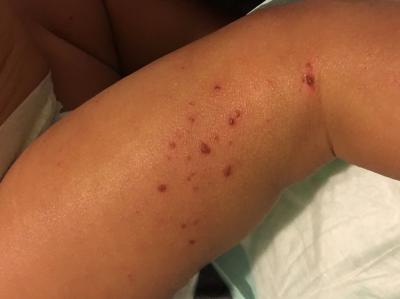
NIAID scientists have found an association between widely used chemicals called diisocyanates and atopic dermatitis, a chronic inflammatory skin disease commonly known as eczema. The researchers also found through an experiment in mice that when bacteria that normally reside on skin are exposed to isocyanate, a component of diisocyanates, they adapt by stopping production of the oils that skin needs to stay healthy. These findings were recently published in the journal Science Advances.

Eczema affects up to 20% of children and up to 10% of adults in high-income countries. People with eczema experience severe itching, skin redness, oozing from the skin, and scaly rashes, all of which can be painful. A particularly stressful aspect of eczema is its unpredictability, as symptoms can worsen from exposure to multiple substances or without any obvious trigger.

Prior to 1970, the prevalence of eczema in the United States was three to six times lower than it is today. While genetic risk factors are known to play a strong role in the incidence of eczema, the rapidly increasing prevalence of the disease indicates that environmental factors are also important. NIAID researchers led by Ian Myles, M.D., M.P.H, sought to identify environmental pollutants that may be contributing to this increase. Dr. Myles is chief of the Epithelial Research Unit in the NIAID Laboratory of Clinical Immunology and Microbiology.

The investigators compared a database that collates clinic visits for atopic dermatitis with two Environmental Protection Agency resources: the Toxics Release Inventory, which tracks the amounts of certain toxic chemicals that are released into the environment for each U.S. zip code; and the Risk-Screening Environmental Indicators model, which models the route of each chemical release through the environment and the potential human exposure that may result. The scientists identified emissions of diisocyanates as the strongest predictor of local eczema rates.

Diisocyanates have been used since the late 1940s to make polyurethane products, such as rigid and flexible foams, coatings, adhesives, sealants, and elastomers (rubbery materials). Exposure to these products has been shown to increase the risk of eczema, according to the NIAID researchers. The active portion of the diisocyanate molecule, the isocyanate side chain, is also a component of other known eczema triggers, such as wildfires, cigarette smoke, and automobile exhaust from catalytic converters, which became mandatory in the United States in 1975. 

The investigators conducted experiments on the skin of mice to explore how isocyanates might contribute to eczema. Previous research demonstrated that exposing mouse skin to diisocyanates could directly induce eczema, yet the mechanisms were unclear. The current study found that when bacteria that live on healthy skin are exposed to isocyanate, they must adapt to survive. When they adapt, these bacteria shift their metabolism away from making the lipids, or oils, that skin needs to stay healthy. This finding suggests that eczema may be treatable by replacing the modified skin bacteria with healthy bacteria, according to the scientists.

Further research is needed to validate the association between environmental exposure to isocyanate or diisocyanates and eczema and to determine whether the causative mechanisms identified in mice are also found in people.

Dr. Myles and colleagues previously reported the results of a small, early-phase clinical trial that found applying healthy versions of a skin bacterium called Roseomonas mucosa to the skin of people with eczema led to a significant reduction in eczema symptoms that persisted even after the medication was stopped. A subsequent, larger, placebo-controlled clinical trial of R. mucosa strains found that the data failed to meet statistical significance for the primary endpoint of EASI-50 (the proportion of patients with at least a 50% improvement in atopic dermatitis disease severity as measured by the Eczema Area and Severity Index). However, the subgroup of study participants who completed the Phase 2 trial demonstrated sustained, modest, but statistically significant clinical improvements that differed by study site diisocyanate levels.

To facilitate their availability as a potentially beneficial probiotic, NIAID is making the R. mucosa strains available for commercial, non-therapeutic development.

References:

J Zeldin et al. Exposure to isocyanates predicts atopic dermatitis prevalence and disrupts therapeutic pathways in commensal bacteria. Science Advances DOI: 10.1126/sciadv.ade8898 (2023).

SM Langan et al. Atopic dermatitis. The Lancet DOI: 10.1016/S0140-6736(20)31286-1 (2020).

BC Martel et al. Translational animal models of atopic dermatitis for preclinical studies. Yale Journal of Biology and Medicine (2017).

IA Myles et al. Therapeutic responses to Roseomonas mucosa in atopic dermatitis may involve lipid-mediated TNF-related epithelial repair. Science Translational Medicine DOI: 10.1126/scitranslmed.aaz8631 (2020).

Contact Information

Contact the NIAID Media Team.

301-402-1663
niaidnews@niaid.nih.gov

Search NIAID Blog

NIAID-Supported Studies Identify Universal Predictors of Antibody Response to Vaccination

NIAID Now |

When healthy people in the same age range are immunized with the same vaccine, some people’s immune systems make substantially more protective antibodies than others. Scientists want to figure out why some people respond to vaccination better than others.

Investigators have identified predictors of a robust antibody response to a few specific vaccines, such as those for influenza, yellow fever, and hepatitis B. Until recently, however, scientists did not know if there was a common predictor of a potent antibody response to most vaccines. If one does exist, researchers reasoned, perhaps it could guide the development of new strategies to enhance the antibody response to vaccination. This would be especially helpful for infants, older adults, and immunosuppressed individuals, who do not always mount an effective immune response to vaccines.

In pursuit of this goal, researchers associated with the NIAID-funded Human Immunology Project Consortium (HIPC) examined immune responses from the blood of 820 healthy adults ages 18 to 55 years before and after they received a shot of one of 13 different vaccines against 11 different pathogens. These vaccines collectively used six different approaches, or platforms, to generate an immune response. The investigators looked for patterns of genes that were turned on, or expressed, in immune cells before vaccination in people who made high, medium, or low levels of antibodies after vaccination. The scientists found that people who had gene expression patterns associated with inflammation before receiving a shot made the highest level of antibodies. The findings were published in the journal Nature Immunology.

Now, researchers want to figure out how to safely induce this level of inflammation and learn if doing so on the day of immunization helps people generate a stronger antibody response to a pathogen than would be expected otherwise.

A caveat to the findings from this study is that the predictive patterns observed in younger adults did not hold up when the researchers applied them to the small number of study participants ages 50 and older. It’s unclear whether other gene expression patterns would predict the antibody response to vaccines in older adults or in children, who were not included in the study. HIPC-affiliated investigators are seeking an answer to this question.

In related research, NIAID-funded HIPC investigators examined immune responses from the same 820 adults who received one of the 13 different vaccines in the earlier study, but this time, the blood samples were drawn over a period of 21 days after vaccination. The researchers looked for a similar pattern of gene expression among all the participants after immunization that could predict the strength of the antibody response several weeks later. Initially, no common pattern appeared for all 13 vaccines. However, when the investigators adjusted the data to synchronize the timing of expression of specific genes, they did find a predictive pattern. The activation and replication of precursors of antibody-producing B cells predicted a robust antibody response to immunization. These findings, also published in Nature Immunology, indicate that after making a time adjustment, scientists can look at gene expression to accurately forecast the magnitude of an individual’s antibody response.

Notably, these two studies do not account for other aspects of the immune response that may be important for vaccine protection, such as the variety of molecules to which the antibodies bind, the strength of antibody binding, and the T-cell response. 

The published and ongoing research reported in the two studies described here is possible because data from numerous clinical trials of more than a dozen different vaccines was shared, standardized, and pooled to answer broad questions about immune responses to vaccines.

The National Institutes of Health Data Management and Sharing policy requires scientists to describe how they will plan to preserve and publicly share data from research funded by or conducted at NIH. Data from the two studies and publications described here is available at www.ImmPort.org, a NIAID-supported data repository that provides access to NIAID-funded research findings. Adherence to the NIH Data Management and Sharing policy and ease of access to shared data will accelerate the pace of biomedical research, enable the validation of research results, and ensure optimal use of high-value datasets.  

References:

S Fourati et al. Pan-vaccine analysis reveals innate immune endotypes predictive of antibody responses to vaccination. Nature Immunology DOI: 10.1038/s41590-022-01329-5 (2022).

T Hagan et al. Transcriptional atlas of the human immune response to 13 vaccines reveals a common predictor of vaccine-induced antibody responses. Nature Immunology DOI: 10.1038/s41590-022-01328-6 (2022).

Contact Information

Contact the NIAID Media Team.

301-402-1663
niaidnews@niaid.nih.gov

Search NIAID Blog

NIH Study Links Specific Outdoor Air Pollutants to Asthma Attacks in Urban Children

Moderate levels of two outdoor air pollutants, ozone and fine particulate matter, are associated with non-viral asthma attacks in children and adolescents who live in low-income urban areas, a study funded by the National Institutes of Health has found. The study also identifies associations between exposure to the two pollutants and molecular changes in the children’s airways during non-viral asthma attacks, suggesting potential mechanisms for those attacks.

Contact

Submit a Media Request

Contact the NIAID News & Science Writing Branch.

301-402-1663
niaidnews@niaid.nih.gov
All Media Contacts

Newly Discovered Species of Gut Bacteria May Cause Some Cases of Rheumatoid Arthritis

NIAID Now |

Research by NIAID grantees strongly suggests that immune responses to a newly discovered species of gut bacteria may cause some cases of a common autoimmune disease called rheumatoid arthritis, or RA. The findings were recently published in Science Translational Medicine.

In people with RA, their own antibodies and T cells attack their joints, especially in the hands, wrists, and knees. This leads to inflammation, pain, and swelling in the joint lining. Over time, RA progresses to irreversible joint tissue damage, chronic pain, loss of function, and deformities. Scientists do not know what causes RA despite extensive efforts to understand the earliest stages of this disease.  

Some RA research has focused on understanding what triggers the development of the antibodies and T cells that attack the joints. Several studies have suggested that the production of joint-targeting antibodies starts at mucosal surfaces, such as the lining of the mouth, airways, and gut, more than a decade before symptoms arise and might be stimulated by bacteria at these sites. Until now, however, the bacterial culprit remained unknown.

In the new study, investigators isolated antibody-secreting cells called plasmablasts from the blood of people at risk for RA and found that the antibodies produced by these cells recognized certain bacteria in the gut microbiome of these individuals. The researchers identified a previously unknown bacterial species targeted by these antibodies, which they named Subdoligranulum didolesgii. This bacterium was present in the feces of four out of 24 people who were either at risk for or diagnosed with RA but absent from the feces of 12 healthy people.

The scientists hypothesized that a mucosal immune response to S. didolesgii in the gut might progress to a body-wide immune response. To test their hypothesis, the researchers gave mice an oral dose of the bacterium and monitored their immune and physical responses. The mice developed antibodies and T cells that attacked their joints and led to visible joint swelling. In addition, the immunologic changes that the scientists observed in the mice extended over time from the gut mucosa to sites throughout the body. 

Taken together, the findings suggest that in some people with RA, immune responses to S. didolesgii in the gut may trigger the production of antibodies and T cells that circulate throughout the body and attack the joints, leading to chronic inflammation. Thus, it appears that RA may develop because immune-system targets shared by S. didoglesgii and joint tissue have a similar structure or amino-acid sequence.

Didolesgi is the Cherokee word for arthritis or rheumatism and was proposed as the name for the newly discovered bacterium by the study’s first author, Meagan E. Chriswell, B.S., who is an enrolled member of the Cherokee Nation of Oklahoma. Ms. Chriswell, an M.D./Ph.D. candidate in immunology at the University of Colorado Anschutz Medical Campus in Aurora, and Kristine A. Kuhn, M.D., Ph.D., an associate professor of medicine at the university, led the research. 

The study was co-funded by NIAID and the National Institute of Arthritis and Musculoskeletal and Skin Diseases, both part of the National Institutes of Health.

Reference: ME Chriswell, et al. Clonal IgA and IgG autoantibodies from individuals at risk for rheumatoid arthritis identify an arthritogenic strain of Subdoligranulum. Science Translational Medicine DOI: 10.1126/scitranslmed.abn5166 (2022).

Contact Information

Contact the NIAID Media Team.

301-402-1663
niaidnews@niaid.nih.gov

Search NIAID Blog

Vaccine Adjuvant Research Programs

Areas of Adjuvant Research

A scientist looking through a microscope

Adjuvant Discovery and Mechanistic Research

Discovery and Mechanistic Research seeks to identify novel adjuvant candidates that can be used to augment the efficacy of human vaccines and elucidate the immune mechanism of novel receptors.

heat map

Adjuvant Comparison

Adjuvant Comparison programs support the side-by-side comparison of experimental vaccines formulated with different adjuvants, to include comprehensive assessments of the strength, quality, breadth, and durability of immune responses.

Vile of vaccines

Adjuvant Development

Adjuvant Development supports the preclinical development of novel adjuvants for use in vaccines targeting immune-mediated or infectious diseases. 

A clinical trial participant getting a vaccine

Clinical Evaluation

This program is intended to address the limited availability of adjuvants that mimic those with a favorable clinical track record or show high potential in late pre-clinical testing.

Contact Information

Points of contact for each program can be found on individual program pages.

Vaccine Adjuvant Compendium (VAC)

The Vaccine Adjuvant Compendium (VAC) was launched in 2021 by the NIAID Division of Allergy, Immunology, and Transplantation (DAIT) to foster collaborations between NIAID-supported adjuvant researchers and the broader scientific community.

Adjuvant Webinar Series

The NIAID Adjuvant Webinar Series presents current research relating to vaccine adjuvants on the fourth Tuesday of every month from 12:30 – 1:30 p.m. (ET) .

Content Coordinator
Content Manager
Banner (Hero) Image
Introduction

NIAID plays a leading role in the discovery, development, and characterization of new vaccine adjuvants that may be used to: improve the efficacy of current vaccines; design new or improved vaccines for existing and emerging infectious diseases; and develop vaccines to treat allergies, autoimmune diseases, and cancer.

Networks & Programs

Injectable Antiretroviral Medication Proves to be Effective in Preventing HIV Infection in Women

NIAID Now |

Women are disproportionately affected by HIV, with young women in sub-Saharan Africa three times as likely to be infected than young men in the same age group. In 2015, the World Health Organization (WHO) recommended that tenofovir-based pre-exposure prophylaxis (PrEP) be offered to high-risk individuals for prevention. As a result, oral PrEP has been introduced in more than 70 countries, including sub-Saharan Africa. Despite expanded access to oral PrEP in the region, women experience challenges to daily pill-taking, including discrimination, difficulties swallowing pills, and the fear of violence from partners, family, and community members. Additionally, oral PrEP drug concentrations in vaginal tissues are lower than in rectal tissues, therefore women must take 6-7 doses per week to achieve levels associated with HIV prevention. An effective alternative to daily PrEP is critical to women’s health and ending the HIV epidemic. 

This phase 3 clinical trial compared the safety and effectiveness of two treatment options for HIV prevention in HIV-uninfected women—daily oral tenofovir diphosphate plus emtricitabine (TDF-FTC) and injectable cabotegravir given every 8 weeks. The study was conducted in 20 clinical sites in seven sub-Saharan African countries where the burden of HIV is high. High-risk women aged 18-45 years were eligible for this study if they agreed to use contraception during the study and reported two vaginal intercourse episodes 30 days prior to enrollment. Study participants were randomized to either of the treatment groups at enrollment and provided adherence counselling, HIV testing, and physical exams at every visit. The clinical trial design ensured that neither the participants nor the study staff could identify which treatment group each participant was assigned to.  

The results of this large trial showed that injectable cabotegravir was superior to daily oral TDF-FTC for the prevention of HIV in women at substantial risk in sub-Saharan Africa. Participants in the cabotegravir group had an 88% lower risk of HIV infection compared with those in the TDF-FTC group. Furthermore, there was no difference in the frequency of adverse events between the two study groups outside of injection site reactions, suggesting cabotegravir was generally safe and well-tolerated. Of particular note, cabotegravir offered an adherence advantage over the daily pills. Most of the participants that acquired a HIV infection in the daily PrEP treatment group had unquantifiable drug concentrations at the time of infection. This study provides evidence that injectable cabotegravir could be a viable alternative to daily oral PrEP for HIV prevention in women in high-incidence settings. In a related study, injectable cabotegravir was shown to be safe and effective in preventing HIV in cisgender and transgender women who have sex with men.  

Reference: Delany-Moretlwe et al. Cabotegravir for the prevention of HIV-1 in Women:  results from HPTN 084, a phase 3, randomized clinical trial, Lancet, April 1, 2022 - Volume 399 – Issue103378 - p 1779-1789.

Contact Information

Contact the NIAID Media Team.

301-402-1663
niaidnews@niaid.nih.gov

Search NIAID Blog